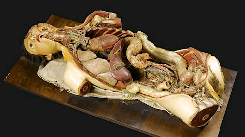
Disturbingly Detailed 17th Century Wax Anatomy Models Go On Display!
Posted by Tony on October 20th, 2012Long before we were able to see inside people with x-rays, tiny cameras or accurate 3D models doctors still had to know what pieces we were made up and where they went. Students learning anatomy didn’t always have the luxury of a bunch of fresh cadavers to study either.
Enter the wax anatomy model.
During the 17th century, there wasn’t any way to learn anatomy unless someone died and their body was immediately trucked-in fresh for people in disciplines that needed to study anatomy. Instead, artists began creating anatomy models out of wax. The intricacy of these models is unsettling and creepy but also amazing because of the stunning extent of the details. Many of these wax models featured things like a removable chest, face or vital organs which, when removed, would reveal even more gruesome details of our inner anatomy.
A lengthy but disturbingly interesting write-up about the details of these models, including photos, was posted on the Journal of Anatomy way back in 2009. Why are we talking about them just now? Well they’re currently on display at the Museum of London.
Often called ‘Venuses’, referencing the Venus Di Medici statue created by an unknown Greek sculptor, most of the models were female forms and several were put in often nightmare-inducing poses.
What do we mean about nightmare-inducing? How about a pregnant woman showing her womb in operation by pulling back flaps of skin on her belly.
We warned you.
[IO9]

October 22nd, 2012 at 2:07 am
Now we’re at the intersection between anatomists and serial killers. The creepy German guy who invented the body plastinization process, he could have been Ed Gein in another life.